English
光超音波イメージングと血管解析
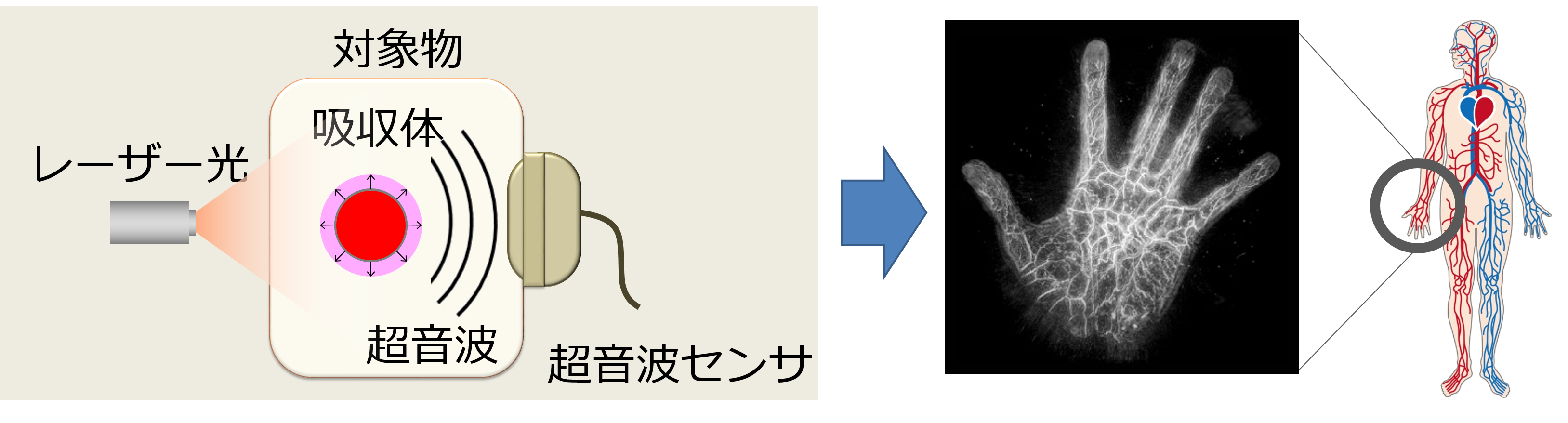
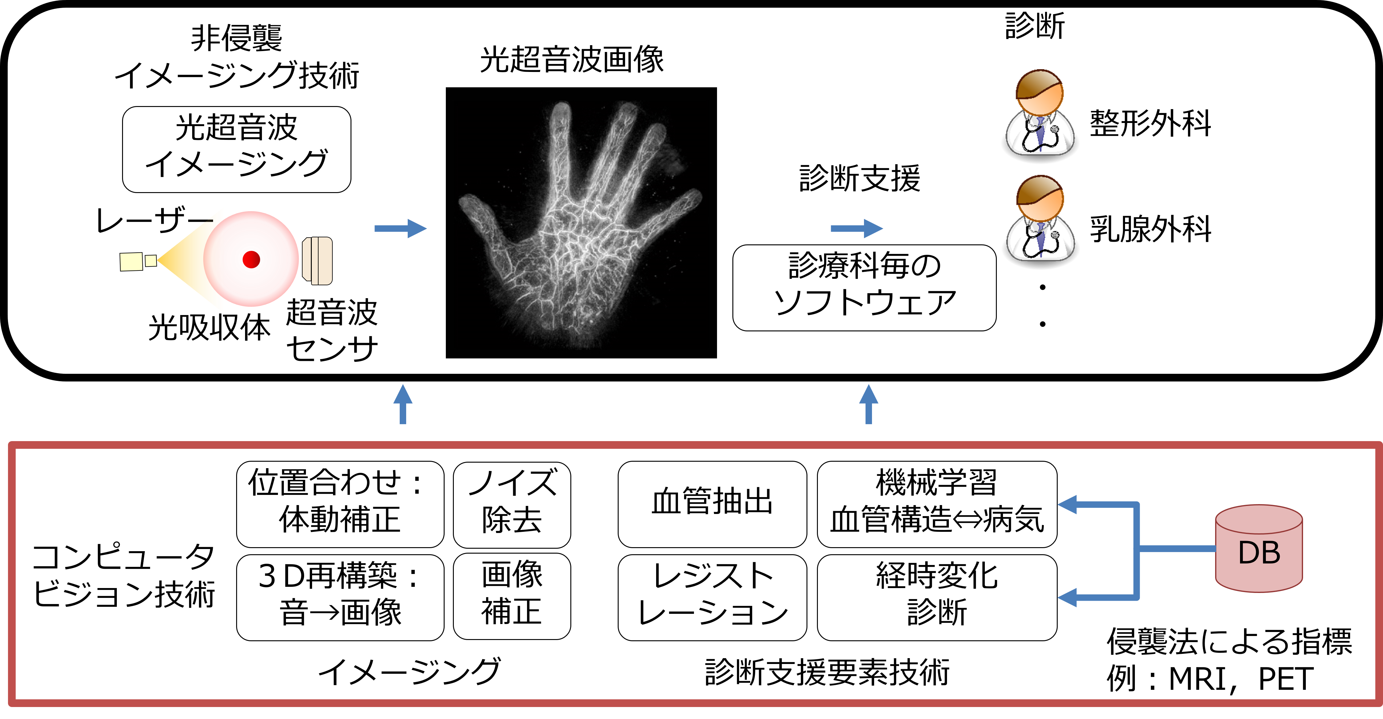
高齢化社会の到来に伴い、健康長寿で豊かな生活を実現し、病気や介護への不安を解消させる技術サポートが求められています。NIIは、病気の早期診断や超精密検査の実現を目指すImPACTに参加し、生体や物体内部を非侵襲・非破壊でリアルタイム三次元可視化する光超音波イメージングの高度化を行っています。光超音波システムは、レーザー照射により発生する超音波を検出し可視化する最先端計測技術です。この技術は、非侵襲・非破壊である上に、透過して深部まで照射できる光と超音波の両方の特性を活かし、肉眼では見えない様々な対象の可視化を可能にします。本研究では、コンピュータビジョン技術により、鮮明な画像を得るイメージング技術の高度化や、様々な情報を用いた画像解析による診断支援を行っています。例えば、撮影中の患者の体動による画質劣化に対して、画像の位置合わせにより患者の動きを補正し、画質改善した診断しやすい画像を提供できるようになります。また、疾病に関係が深い血管状態を把握するため、血管構造の自動抽出技術の開発を進めています。
Semi-Supervised Learning with Structured Knowledge for Body Hair Detection in Photoacoustic Image
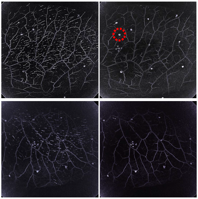
Photoacoustic (PA) imaging is a promising new imaging technology for non-invasively visualizing blood vessels inside biological tissues. In addition to blood vessels, body hairs are also visualized in PA imaging, and the body hair signals degrade the visibility of blood vessels. For learning a body hair classifier, the amount of real training and test data is limited, because PA imaging is a new modality. To address this problem, we propose a novel semi-supervised learning (SSL) method for extracting body hairs. The method effectively learns the discriminative model from small labeled training data and small unlabeled test data by introducing prior knowledge, of the orientation similarity among adjacent body hairs, into SSL. Experimental results using real PA data demonstrate that the proposed approach is effective for extracting body hairs as compared with several baseline methods.
Digital Artery Deformation on Movement of The Proximal Interphalangeal Joint
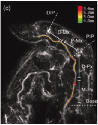
This study aimed to characterize in vivo human digital arteries in three-dimensions using photoacoustic tomography in order to understand the specific mechanism underlying arterial deformation associated with movement of the proximal interphalangeal joint. Three-dimensional morphological data were obtained on the radialis indicis artery (radial artery of the index finger) at different angles of the joint. The association between increased curvature of the deformation and the anatomical region was assessed. Characteristic morphological deformations in areas of major deformation were determined. The deformation of the artery was characterized by three consecutive curves in juxta-articular regions, which were particularly noticeable when the joint was flexed at an angle of???60°. The change in the curvature of the deformation during 30°?90° of flexion was lower in middle-aged individuals than in young individuals. Better understanding of the mechanism underlying deformation of the digital arteries may contribute to advancements in flap procedures and rehabilitation strategies after digital artery repair.
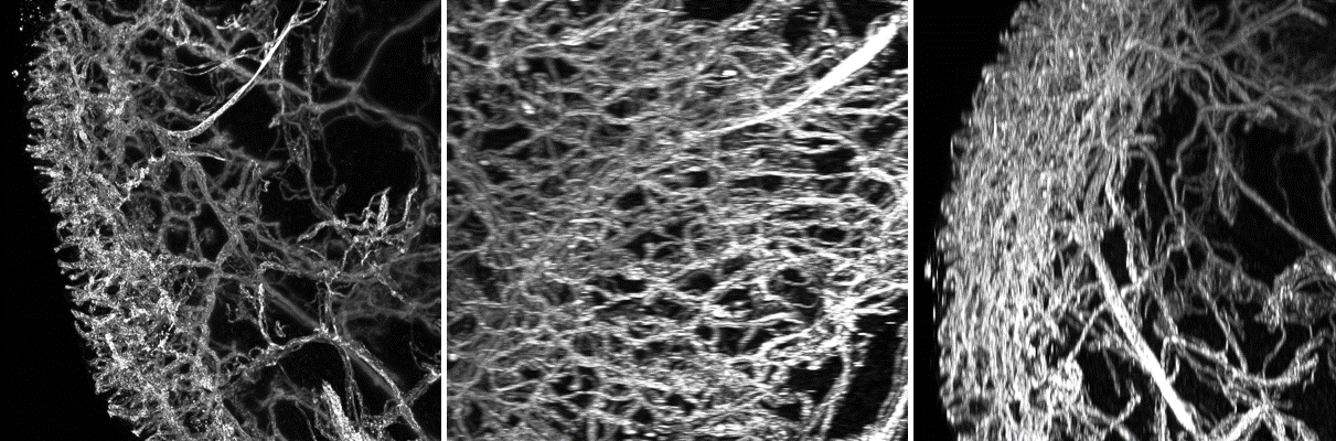
Light-Sheet Microscopy Reveals Site-Specific 3-Dimensional Patterns of the Cutaneous Vasculature and Pronounced Rarefication in Aged Skin
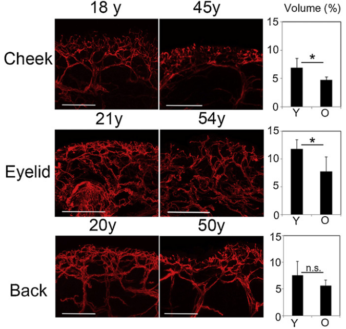
The skin has a 3-dimensional structure that includes several appendages, such as sweat glands and sebaceous glands, as well as lymphatic and blood vessels and nerves. In spite of its complex three-dimensional structure, histological studies of the skin, including quantitative analyses of specific cell types and structures, predominantly use two-dimensional sections. Thus, for example the tortuosity of a blood vessel is not captured in histological sections. We next analyzed the pattern of cutaneous capillaries in different areas of the skin, namely the cheek, eyelid and back skin, and investigated whether there might occur changes during aging. First, the dermal area was divided into an upper and lower area and morphometric 3-dimensional analyses, including determination of the relative volume, diameter and number of branching points of capillaries were performed in an upper dermal area of 300 mm-depth below the epidermis. We found that eyelid skin has the densest capillary network below the epidermal layer, as compared to the cheek skin and the back skin
Virtual Blood Vessels using Stereo X-ray Images
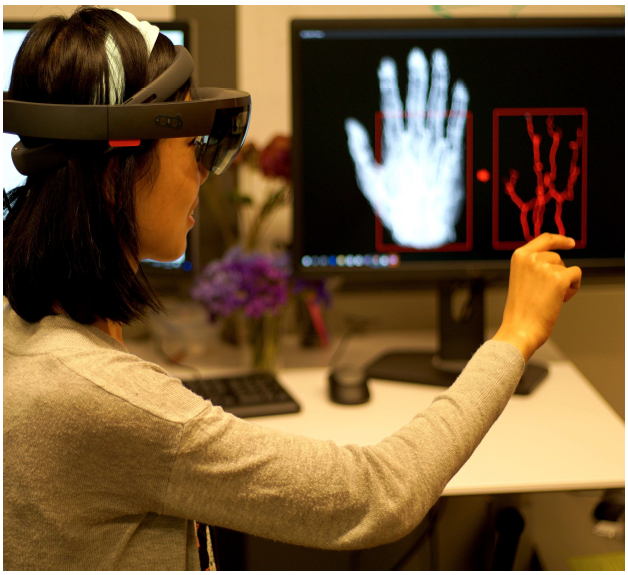
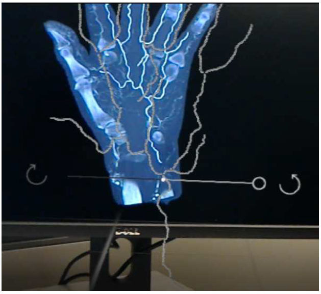
We propose a fully automatic system to reconstruct and visualize 3D blood vessels in Augmented Reality (AR) system from stereo X-ray images with bones and body fat. Currently, typical 3D imaging technologies are expensive and carrying the risk of irradiation exposure. To reduce the potential harm, we only need to take two X-ray images before visualizing the vessels. Our system can effectively reconstruct and visualize vessels in following steps. We first conduct initial segmentation using Markov Random Field and then refine segmentation in an entropy based post-process. We parse the segmented vessels by extracting their centerlines and generating trees. We propose a coarse-to-fine scheme for stereo matching, including initial matching using affine transform and dense matching using Hungarian algorithm guided by Gaussian regression. Finally, we render and visualize the reconstructed model in a HoloLens based AR system, which can essentially change the way of visualizing medical data. We have evaluated its performance by using synthetic and real stereo X-ray images, and achieved satisfactory quantitative and qualitative results.
Blood Vessel Extraction by Semi-Supervised Learning
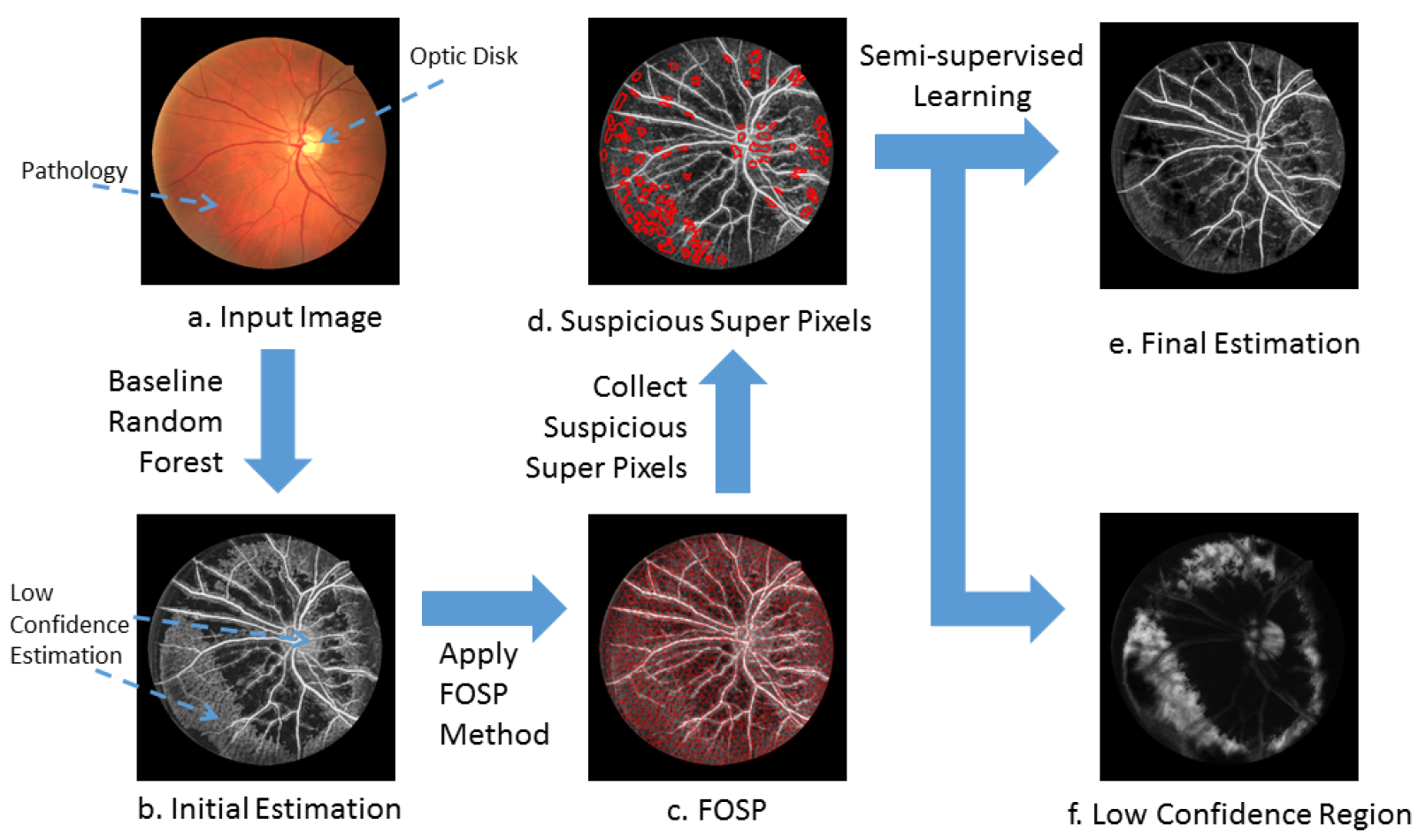
In this paper, we focus on semi-supervised learning for biomedical image segmentation, so as to take advantage of huge unlabelled data. We observe that there usually exist some homogeneous connected areas of low confidence in biomedical images, which tend to confuse the classifier trained with limited labelled samples. To cope with this difficulty, we propose to construct forest oriented super pixels(voxels) to augment the standard random forest classifier, in which super pixels(voxels) are built upon the forest based code. Compared to the state-of-the-art, our proposed method shows superior segmentation performance on challenging 2D/3D biomedical images. The full implementation (based on Matlab) is available at https://github.com/lingucv/ssl_superpixels.
Separation of Transmitted Light and Scattering Components in Transmitted Microscopy
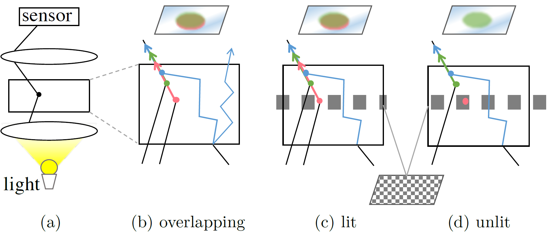
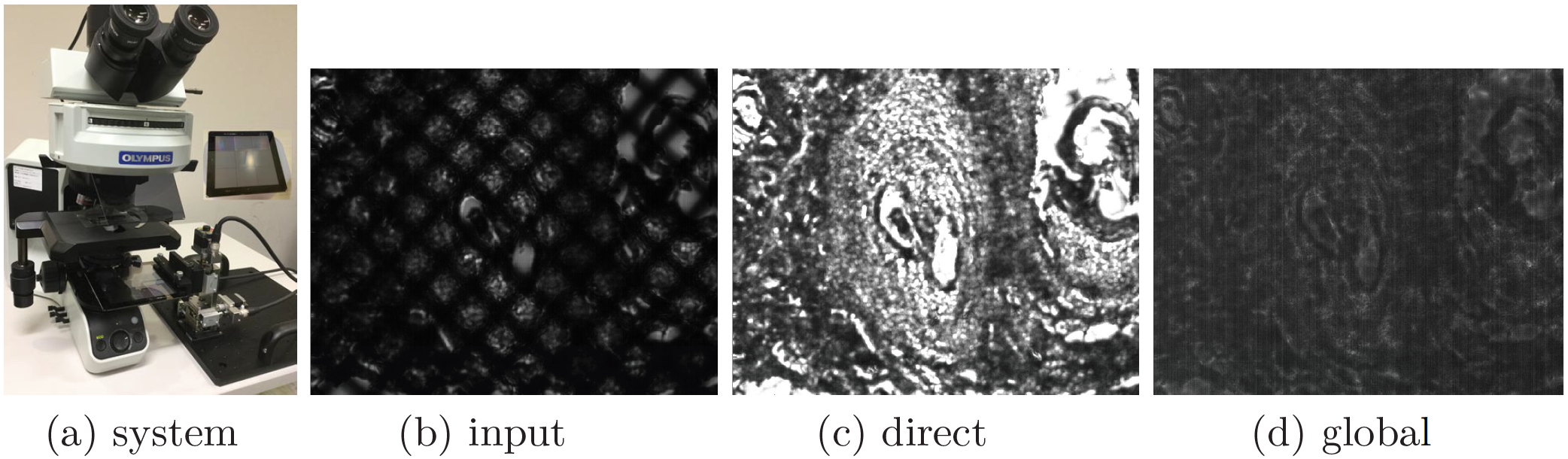
In transmitted light microscopy, a specimen tends to be observed as unclear. This is caused by a phenomenon that an image sensor captures the sum of these scattered light rays traveled from different paths due to scattering. To cope with this problem, we propose a novel computational photography approach for separating directly transmitted light from the scattering light in a transmitted light microscope by using high-frequency lighting. We first investigated light paths and clarified what types of light overlap in transmitted light microscopy. The scattered light can be simply represented and removed by using the difference in observations between focused and unfocused conditions, where the high-frequency illumination becomes homogeneous. Our method makes a novel spatial multiple-spectral absorption analysis possible, which requires absorption coefficients to be measured in each spectrum at each position. Experiments on real biological tissues demonstrated the effectiveness of our method.
Vascular Registration in Photoacosutic Imaging by Low-Rank Alignment
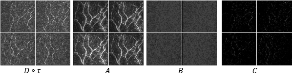
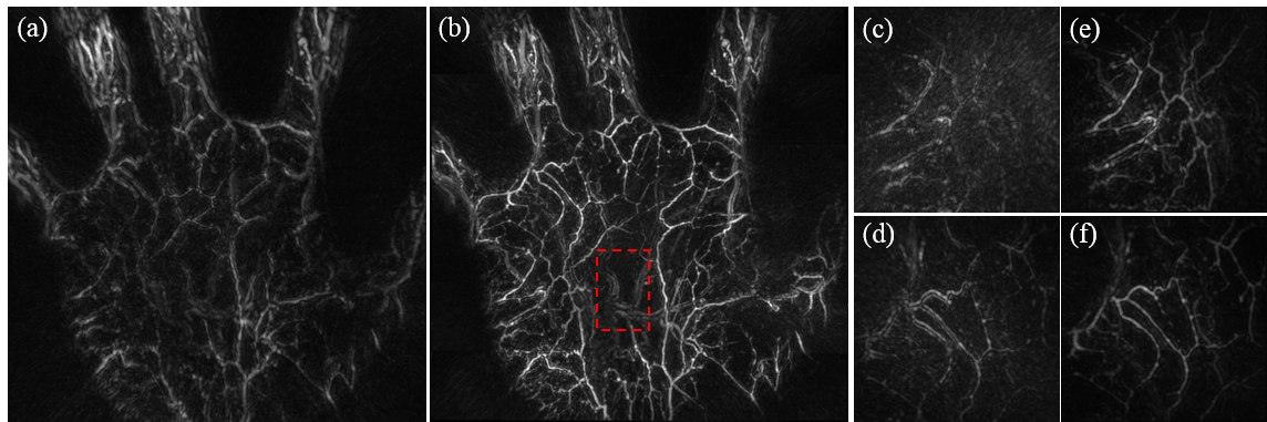
Photoacoustic (PA) imaging has been gaining attention as a new imaging modality that can non-invasively visualize blood vessels inside biological tissues. In the process of imaging large body parts through multi-scan fusion, alignment turns out to be an important issue, since body motion degrades image quality. In this paper, we carefully examine the characteristics of PA images and propose a novel registration method that achieves better alignment while effectively decomposing the shot volumes into low-rank foreground (blood vessels), dense background (noise), and sparse complement (corruption) components on the basis of the PA characteristics. The results of experiments using a challenging real data-set demonstrate the efficacy of the proposed method, which significantly improved image quality, and had the best alignment accuracy among the state-of-the-art methods tested.
3D Structure Modeling of Dense Capillaries by Tracking
A newly developed imaging technique called light-sheet laser microscopy imaging can visualize the detailed 3D structures of capillaries. Capillaries form complicated network structures in the obtained data, and this makes it difficult to model vessel structures by existing methods that implicitly assume simple tree structures for blood vessels. To cope with such dense capillaries with network structures, we propose to track the flow of blood vessels along a base-axis using a multiple-object tracking framework. We first track multiple blood vessels in cross-sectional images along a single axis to make the trajectories of blood vessels, and then connect these blood vessels to reveal their entire structures. This framework is efficient to track densely distributed vessels since it uses only a single cross-sectional plane. The network structure is then generated in the post-processing by connecting blood vessels on the basis of orientations of the trajectories. The results of experiments using a challenging real data-set demonstrate the efficacy of the proposed method, which are capable of modeling dense capillaries.